DermaDiagnosis

As Problem Spreads, Man Seeks Help

Author and Disclosure Information


For several months, a 30-year-old man has had an asymptomatic rash on his legs. The lesions first appeared on his lower legs and ankles; over the subsequent months, they have spread upward. Now, the rash reaches to just below his knees. During this time, he has had two bouts of strep throat, both adequately treated. He denies any other skin problems and has no relevant family history. The patient denies alcohol or drug abuse and is not taking any prescription medications. Prior to referral to dermatology, he was seen in two urgent care clinics; at one, he received a diagnosis of fungal infection and at the other, of “vitamin deficiency.” He was given a month-long course of terbinafine (250 mg/d) that produced no change in his rash. He achieved the same (non)result from an increased intake of vitamins. Examination reveals annular reddish brown macules, measuring 1 to 3 cm, sparsely distributed from the knees to just above the ankles on both legs. The lesions are a bit more densely arrayed on the anterior legs. There is no palpable component to any of them and no discernable surface scale. Digital pressure fails to blanch the lesions. The hairs and follicles on the patient’s legs appear normal. There are no notable skin changes elsewhere, and the patient is alert, oriented, and in no distress.

Given the facts of the case as presented, the most likely diagnosis is

a) Scurvy

b) Schamberg disease

c) Cutaneous T-cell lymphoma

d) Thrombocytopenia

ANSWER
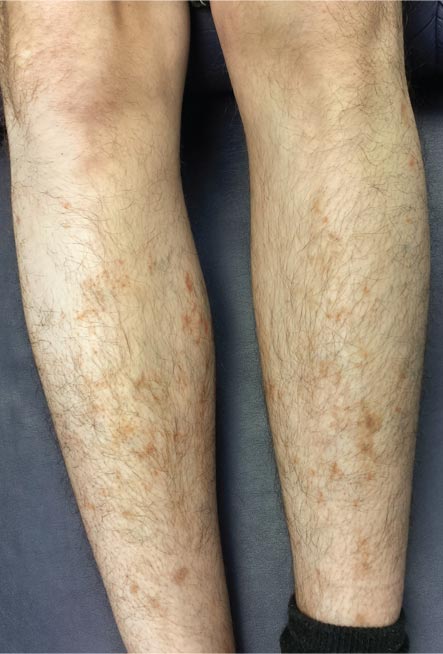
The correct answer is Schamberg disease (choice “b”), a benign form of capillaritis; see Discussion for more information.

Scurvy patients can present with ecchymosis (among other findings that were missing in this case). But scurvy (choice “a”) is rare, and by the time the disease is evident, the patient is typically quite ill.

Cutaneous T-cell lymphoma (choice “c”) can manifest as purpuric annular lesions. However, these would be unlikely to take the distributive pattern seen in this case, and they usually have an atrophic surface.

Thrombocytopenia (choice “d”) and other coagulopathies, although rightly considered, would probably manifest in other ways as well (ie, not just cutaneously).

Continue for the discussion >>

Pages

Recommended Reading

Common Presentation for Complex Condition
Clinician Reviews
FDA Panel Backs Injectable Treatment For Chin Fat
Clinician Reviews
ACP Guidelines For Preventing, Treating Pressure Ulcers
Clinician Reviews
Halting Biologics Before Surgery Tied to Flares in Psoriasis, Psoriatic Arthritis
Clinician Reviews
VIDEO: Once-yearly Skin Cancer Screening Might Not Be Enough After Organ Transplants
Clinician Reviews
Methotrexate and Biologics Linked to Higher Zoster Risk in Psoriasis
Clinician Reviews
Pimecrolimus Cream Safe, Effective for Atopic Dermatitis in Young Children
Clinician Reviews
VIDEO: Sun Protection Urged for Asian, Hispanic Women
Clinician Reviews
Attempt at “Wart” Removal Backfires
Clinician Reviews
VIDEO: PDT Tops Blue-light Therapy for Actinic Keratosis
Clinician Reviews